
Overview
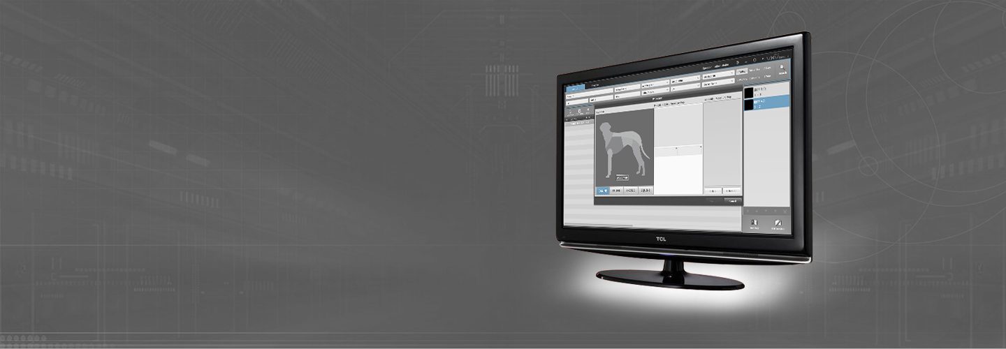
SuperXvue® D-XR is our own developed digital X-ray image acquisition and processing software for vets. This professional acquisition software is designed for X-ray images generated by X-ray detector systems (DR). It also controls the operation of X-ray generators and X-ray units, thus ensuring an efficient and orderly workflow. The user-friendly and straightforward modern graphical user interface (GUI) via touchscreen and mouse. The program was designed, developed and tested in cooperation with medical practitioners in order to provide a user-friendly tool for everyday veterinary care.
Daily work routines are made easier by an intuitive design and an array of integrated functions. This includes a multimedia X-ray positioning guide as well as special filters for optimal bone and soft tissue imaging as well as measurement tools for HD, TPLO, TTA, MMP distraction index and cardiac traits (e.g., VD Thorax Measurement and VHS).
SuperXvue® D-XR software can be integrated with existing information management systems. Furthermore, X-ray images can be evaluated using the SuperXvue® D-XR viewer module included in the acquisition software. Thus, the system functions as a diagnostic workstation.


Benefits of SuperXvue® D-XR Software
- Modern graphical user interface (GUI) adaptable to your language
- Touchscreen operation – ensures a quick, efficient and orderly workflow
- Patient data are captured via DICOM Worklist, BDT/GDT, HL7 or other protocols – data can also be captured manually
- DICOM procedure codes are used to transfer all records relevant to an examination directly from associated information management systems (e.g., HIS/RIS)
- Being available for 200 or more projections(can be customized) and a multitude of adjustment parameters to facilitate visualizing images of animal body parts stored in the system
- Safe and quick registration of emergency patients
- Scheduled examination order can be revised post factum in order to avoid frequent patient repositioning
- Images can be appended to records even after examinations are complete
- Fully integrated radiographic positioning guide for all examinations in veterinary medicine ( can be customized with comprehensive notes, photographs, and sample X-ray images)
- AEC (Automatic Exposure Control) and ARP (Anatomical Programmed Radiography) automatically set X-ray parameter values for each exposure; manual adjustments can easily be made
- Electronic X-ray examination log
- Perfect images every time – few adjustments needed
- Integrated software for automatic image optimization
- Professional image processing – adaptable to different types of examinations according to customer demands
- Special image processing procedures ensure optimal images over a wide range of X-ray parameter settings (e.g., dose reduction)
- Bones and soft tissue can be examined in one image for a better diagnosis
- Details of bones and microstructures are very easy to recognize
- Noise reduction
- Black mask (automatic shutters)
- Automatic removal of grid lines when using fixed grids
- AIAA (automatic image area analysis): Automatically analyses each image for soft tissue and bone structures and applies the most suitable image processing algorithms
- MFLA (multi frequency level analysis): Analyses each image on various frequency levels for ideal sharpness and high subtle contrast
- ANF (automatic noise filter): Algorithm for optimal noise reduction
- GLI (gridless imaging): Exposures without grid. Enables the display of an image as if it had been taken with a grid
- AGLS (automatic grid line suppression): Automatically removes gridlines from flat panel images
- IBC (intelligent brightness control): Automatically displays the image at the ideal level of brightness
- ACO (automatic contrast optimization): Automatic contrast equalization across the entire image – this enables the optimal display of soft tissue and bones at the same time
- ABBS (automatic black border shutter): Automatically darkens all parts of an image outside the collimated area – varying degrees of transparency are available and manual adjustments are easy to make.
- Integrated SuperXvue® D-XR image diagnosis, processing, and storage in an SQL database, including image manipulation, export options, layout adjustments, user interface customization and much more…
- PAN, Steplesszoom, Magnification tool, ROI, Crop, Rotate, Mirror etc.
- Insertion of image annotations, e.g. text, arrow, ellipse
- Adjustment of window/level options as well as gamma correction, sharpening filters and noise suppression.
- Measurement of distances, angles, areas and density
- Special functions for veterinary medicine (e.g., filters for optimal bone and soft tissue imaging, measurement tools for TPLO and TTA, MMP, distraction index and Buchanan‘s Vertebral Heart Score for dogs and cats)
- Many additional functions such as HD measurements, pelvic obliquity measurements, automated generation of medical reports etc.
- Images can be printed with Windows OS printers as well as laser imagers via DICOM Basic Print
- Built-in email tool for image distribution – no external email application necessary
- Image distribution to multiple recipients via image management systems
- Worldwide distribution of images to medical staff and patients via web server, images can be exported in JPEG, PNG, BMP and DICOM formats